Scale Healthcare Machine Learning Through Expert Medical Image Annotation
Machine learning can change how care is delivered, but only when models are trained on data that clinical experts have labeled correctly. CapeStart annotates across ophthalmology, radiology, cardiology and laparoscopy, from single-class bounding boxes through to voxel-level segmentation, and delivers straight into your training pipeline, supported by our broader AI services for clinical data preparation, model validation and AI-assisted imaging.
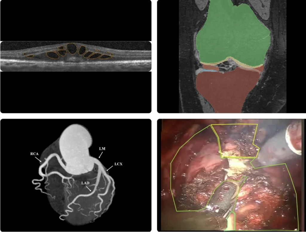
| Project output from our annotation team. | |
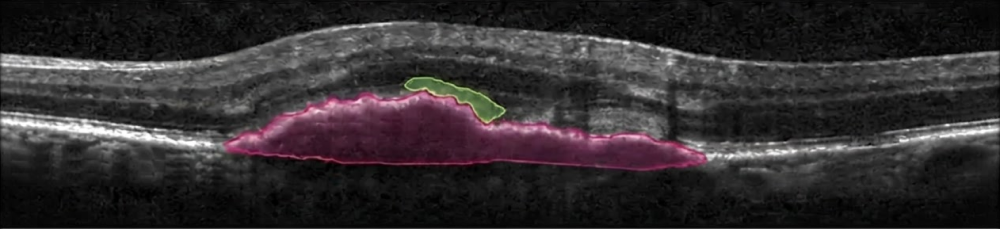
| Top Row | Fluid segmentation on a retinal OCT b-scan, and knee segmentation on MRI. |
| Bottom Row | Coronary vessel labeling on CT angiography, and prostate segmentation on robotic surgical video. |
Four Practices, One Clinical Team
Every sample is project output from our own clinical team, with the modality, the annotation type and the classes named on each one.
Our ophthalmology team annotates anterior and posterior segment images for AI model development, model validation and clinical research.
Anterior segment work covers pterygium, bullous keratopathy and other corneal abnormalities. Posterior segment work covers the features behind grading in diabetic retinopathy, age-related macular degeneration and cystoid macular edema, from lesion detection through to pixel-level segmentation. We also annotate veterinary and preclinical imaging.
IMAGE TYPES AS-OCT, color fundus, OCT, fluorescein angiography, surgical video
ANNOTATION TYPES Segmentation, bounding boxes, boundary tracing, keypoints, polygons
SUPPORTS Model training and validation, clinical research, preclinical and veterinary studies
Top Row: Multi-class lesion annotation on color fundus, and fluid segmentation on a retinal OCT b-scan.
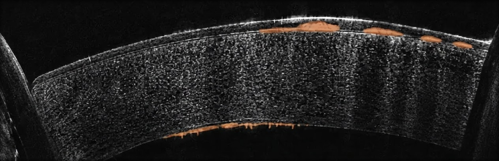
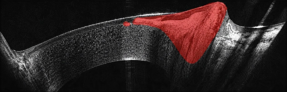
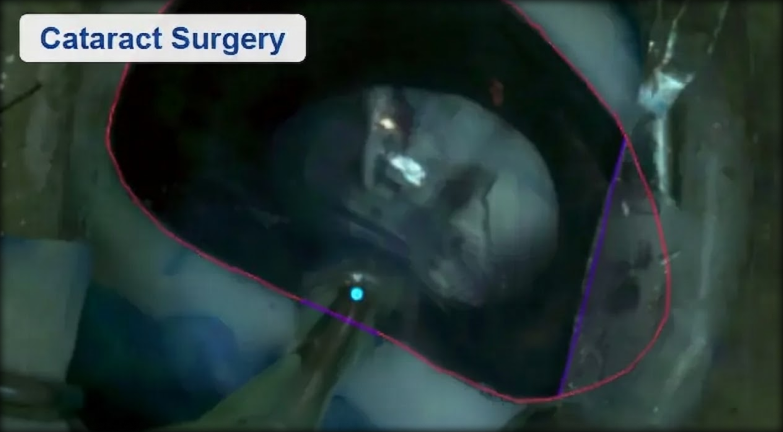
Bottom Row: Corneal pathology on anterior segment OCT, and anatomy and instrument annotation on cataract surgical video
Our radiology annotation team identifies and labels anatomical structures, lesions, abnormalities and clinically relevant findings across six modalities.
CT and MRI work covers multi-organ and voxel-level segmentation, lesion and tumor contouring, the vertebrae, the joints and the soft tissues. Ultrasound covers obstetric, fetal and cardiac studies, mammography covers masses, calcifications and BI-RADS scoring, and X-ray covers abnormality classification, lung findings and Cobb angle measurement, alongside surgical navigation.
IMAGE TYPES MRI, CT, PET/CT, ultrasound, mammography, X-ray
BODY REGIONS Head and neck, brain, chest, abdomen, pelvis, spine and vertebrae, joints, bone, breast
ANNOTATION TYPES 2D and 3D segmentation, bounding boxes, polygonal contours, landmarks, keypoints, measurements
Top Row: Knee segmentation on MRI, and renal mass segmentation on CT.
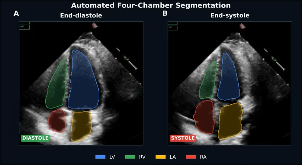
Bottom Row: Four chamber segmentation on fetal ultrasound, and a lesion carried across paired PET and CT.
Annotation of the aortic, mitral, tricuspid and pulmonary valves, including the anatomy of the valves, annulus, leaflets, chambers and vessels, with segmentation, landmarks and measurements for valve planning and interventional procedures.
Modality coverage runs across echocardiography, angiography and cardiac CT and MRI, from coronary vessel labeling on a volume rendered angiogram through to device position on a live fluoroscopic run. We also annotate ECG waveforms, intervals and rhythms, along with EP mapping data.
IMAGE TYPES Coronary CT angiography, echocardiography, fluoroscopy, cardiac CT and MRI, ECG and EP mapping
ANNOTATION TYPES Segmentation, landmarks, measurements, waveform and interval labeling
SUPPORTS Valve planning, interventional procedures, arrhythmia detection
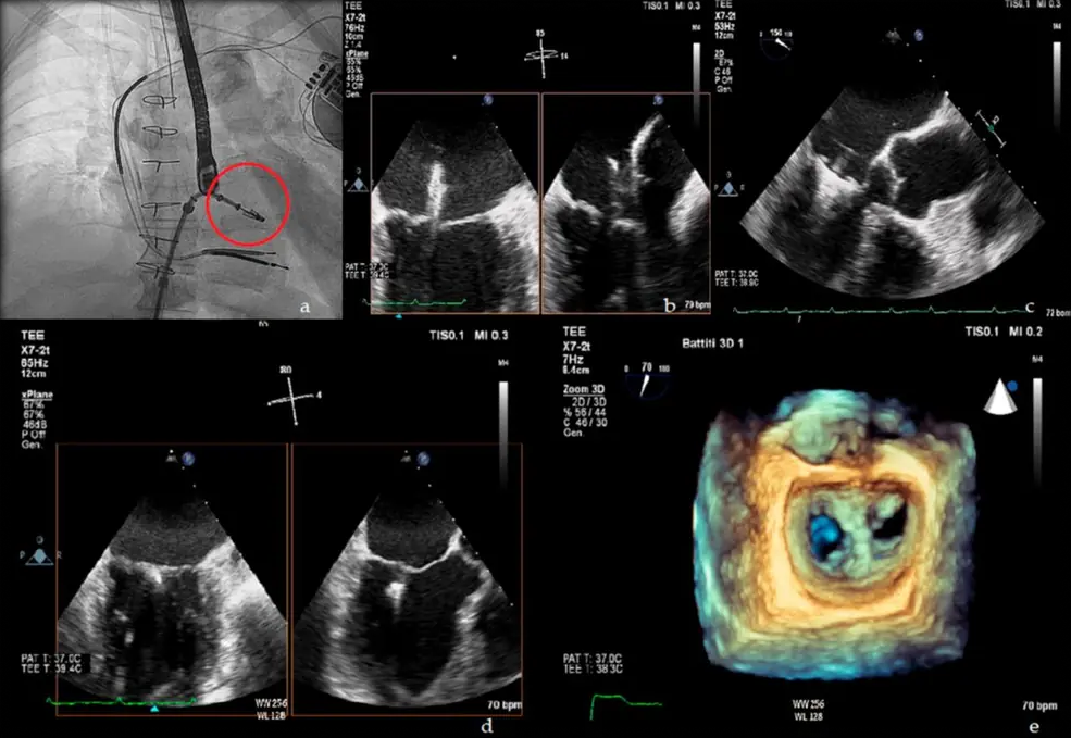
Top Row: Coronary vessel labeling on CT angiography, and one valve study annotated across fluoroscopy, echo and a 3D reconstruction.
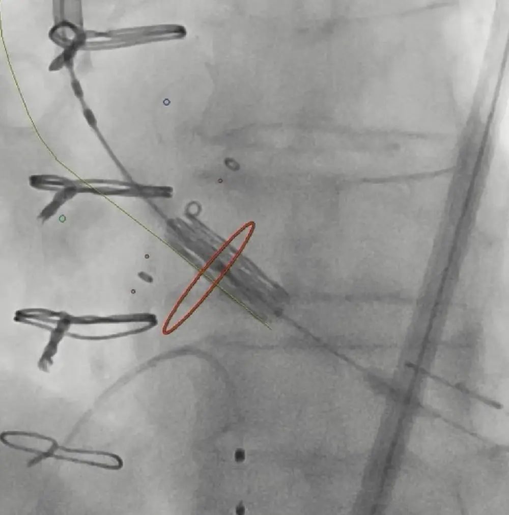
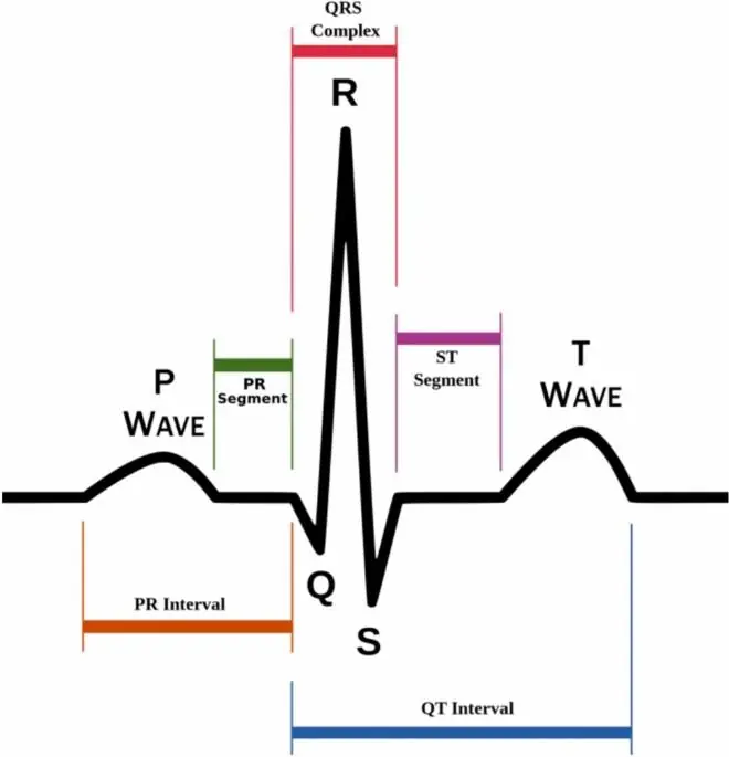
Bottom Row: Device labeling on a fluoroscopic frame, and the ECG waveform and interval scheme.
Anatomical structure identification, surgical instrument detection and tracking, tissue segmentation, surgical phase recognition and procedure assessment.
Anatomy is outlined class by class on both laparoscopic and robotic frames, covering the liver, gallbladder, cystic artery, cystic duct and prostate. Instruments are located, classified and tracked frame to frame, and procedure stages are recognized and assessed across a full operation.
IMAGE TYPES Laparoscopic video, robotic surgical video, still surgical frames
ANNOTATION TYPES Segmentation, polygons, bounding boxes, keypoints, classification, frame-to-frame tracking
SUPPORTS Instrument detection, surgical phase recognition, procedure assessment
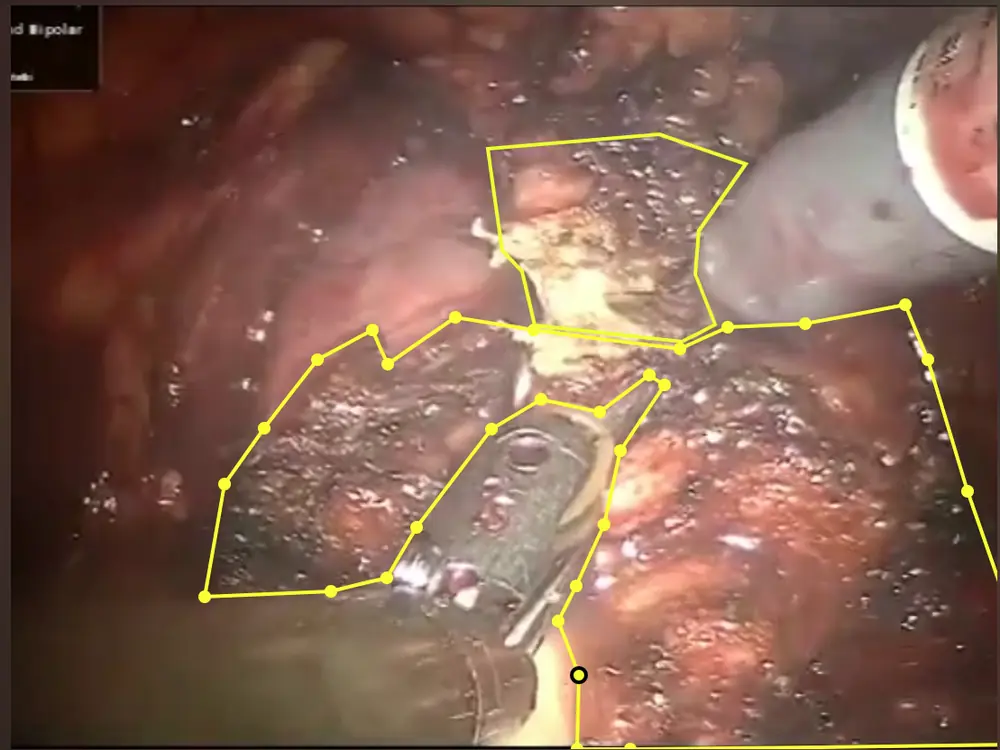
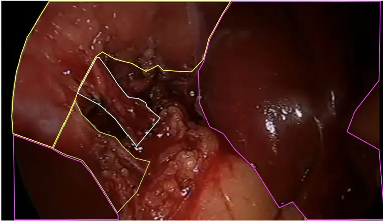
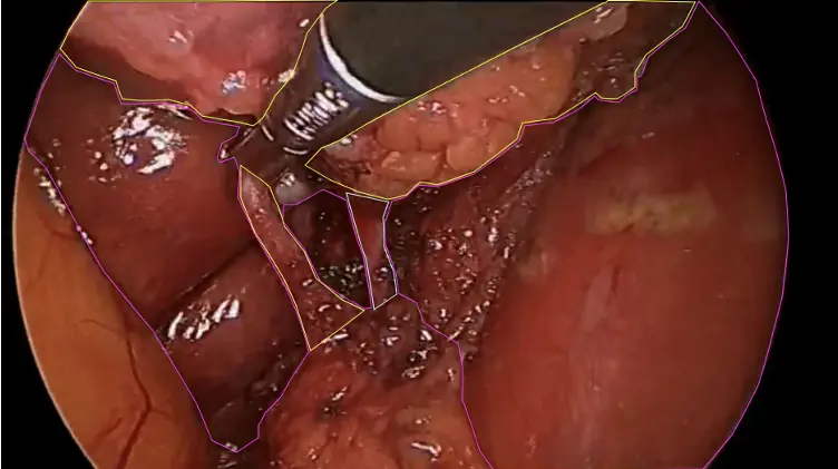
Top Row: Prostate and tissue planes on robotic surgical video, and the hepatobiliary structures on laparoscopic video.
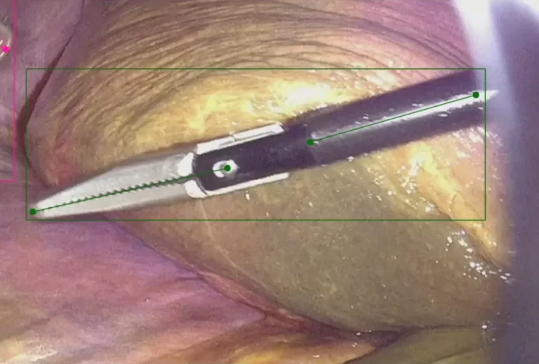
Bottom Row: The same classes carried with the instrument in frame, and instrument detection with keypoints.
Our ophthalmology team annotates anterior and posterior segment images for AI model development, model validation and clinical research.
Anterior segment work covers pterygium, bullous keratopathy and other corneal abnormalities, along with the eyelids and meibomian glands.
Posterior segment work covers the features behind grading in diabetic retinopathy, age-related macular degeneration and cystoid macular edema, from lesion detection through to pixel-level segmentation of fluid, vessels and retinal layers. We also annotate veterinary and preclinical imaging, including porcine cataract surgical video and retinal layers in mice.
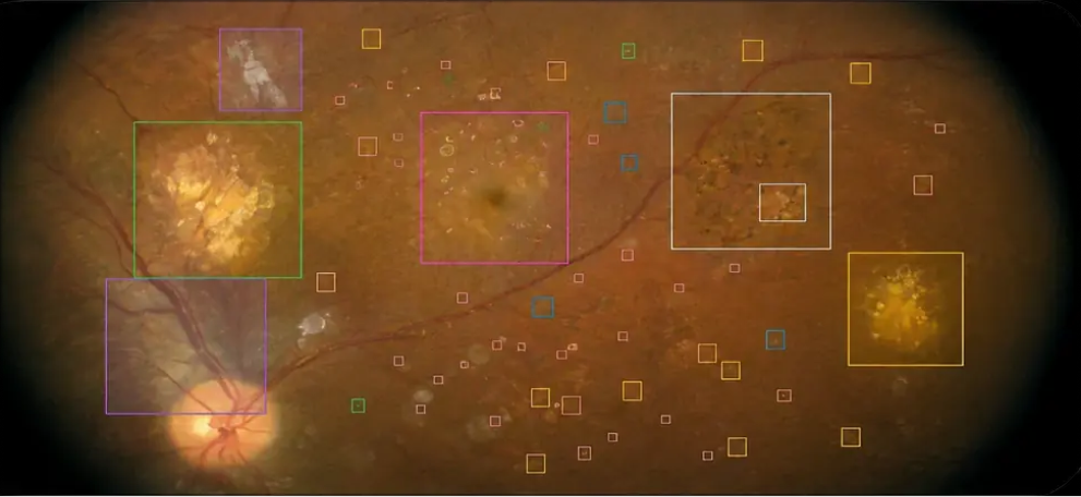
IMAGE TYPES AS-OCT, color fundus, OCT, fluorescein angiography, surgical video
ANNOTATION TYPES Segmentation, bounding boxes, boundary tracing, keypoints, polygons
SUPPORTS Model training and validation, clinical research, preclinical and veterinary studies
Individual lesion classes annotated separately, then carried together on one image.
Every lesion class carried on a single macula-centered image, from large atrophic areas down to individual drusen.
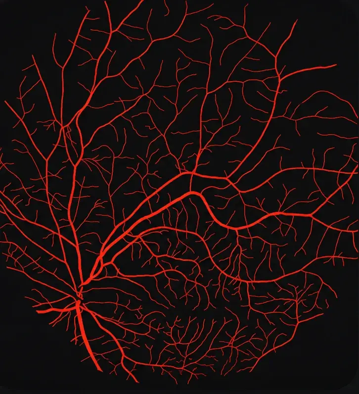
The full vessel tree segmented down to the smallest visible branches, delivered as a binary mask.
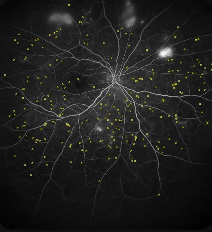
Several hundred microaneurysms marked as individual points across the posterior pole of one eye.
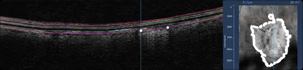
Layer boundaries and fluid compartments, traced in individual b-scans.
Retinal layer boundaries traced across the b-scan, with the lesion outlined on the paired en face image and geographic atrophy measured.
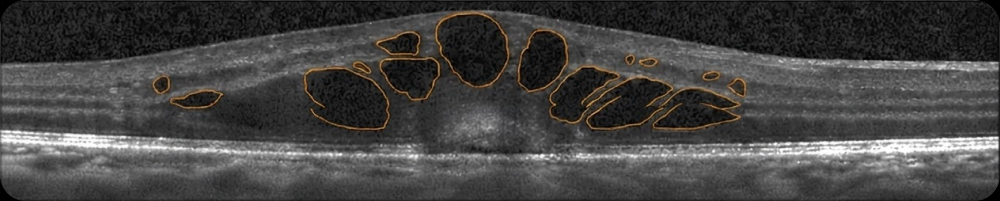
Each cystoid space outlined separately rather than as one region, so cyst count and area are both recoverable. Diffuse retinal thickening is annotated on the same scan.
Subretinal fluid and pigment epithelial detachment segmented as separate classes.
Corneal pathological features, including Bowman's layer scarring and Fuchs' dystrophy, segmented in a single section of the cornea.
Pterygium extent segmented as one region, with a stromal scar anterior to the main body labeled independently.
Annotation supporting device and therapeutic development programs, including surgical video.
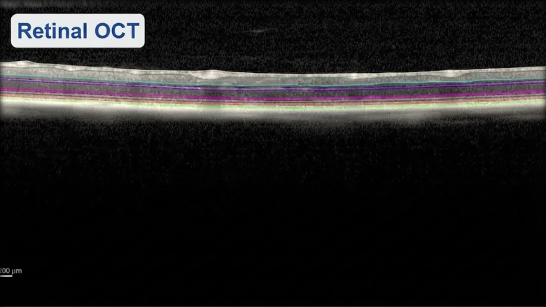
Layer boundaries of mice traced in OCT, where the layers are a fraction of the thickness seen in human scans.
Anatomy outlined, instrument located and the incision point marked on a single surgical frame.
Our radiology annotation team identifies and labels anatomical structures, lesions, abnormalities and clinically relevant findings across six modalities.
CT and MRI work covers multi-organ and voxel-level segmentation, lesion and tumor contouring, the vertebrae, the joints and the soft tissues, with axis measurements where the study calls for them.
Ultrasound covers obstetric and fetal studies, fetal echocardiography and the standard cardiac planes. Mammography covers masses, calcifications, asymmetries and BI-RADS scoring. X-ray covers abnormality classification, lung findings and Cobb angle measurement. Surgical navigation work annotates preoperative and intraoperative studies for real-time guidance.
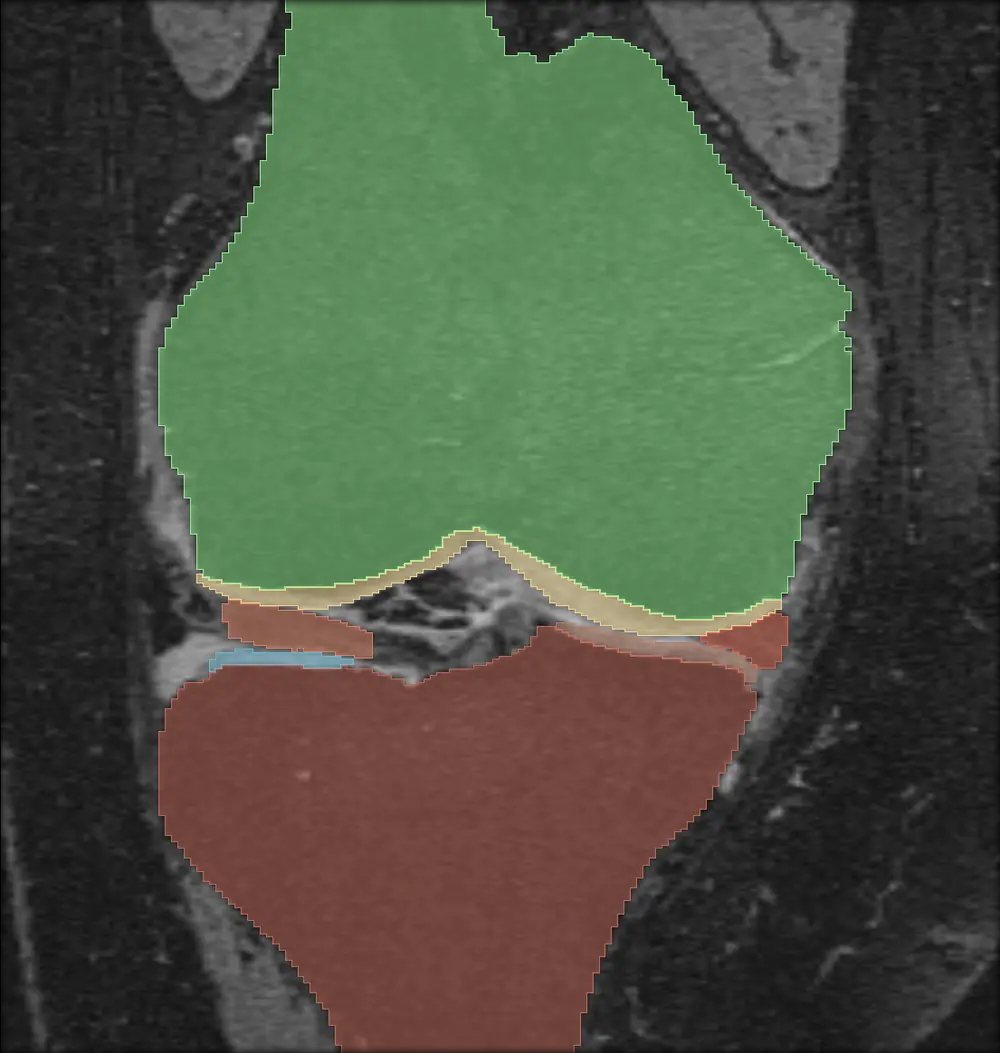
IMAGE TYPES MRI, CT, PET/CT, ultrasound, mammography, X-ray
BODY REGIONS Head and neck, brain, chest, abdomen, pelvis, spine and vertebrae, joints, bone, breast
ANNOTATION TYPES 2D and 3D segmentation, bounding boxes, polygonal contours, landmarks, keypoints, measurements
Voxel-level segmentation of organs, anatomical structures and pathological regions.
Sagittal knee study with the bone, cartilage and ligament structures each segmented as its own class.
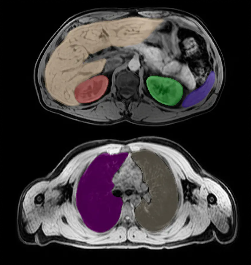
Two slices from one study, with the liver, both kidneys and the spleen segmented on the abdominal slice and the lungs on the chest slice.
Lesions contoured region by region, with axis measurements where the study calls for them.
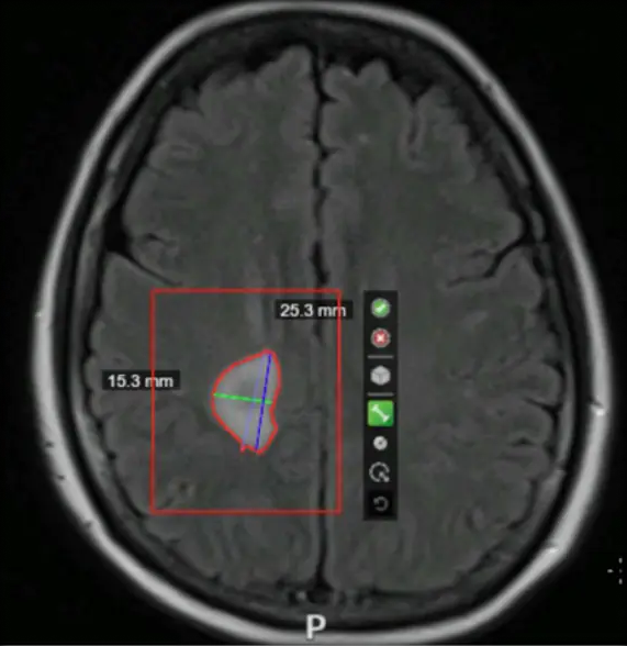
Brain lesion contoured on a FLAIR slice, with long and short axis measurements taken inside the region of interest.
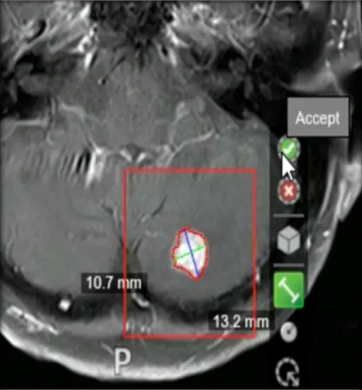
A second brain lesion contoured and measured on both axes.
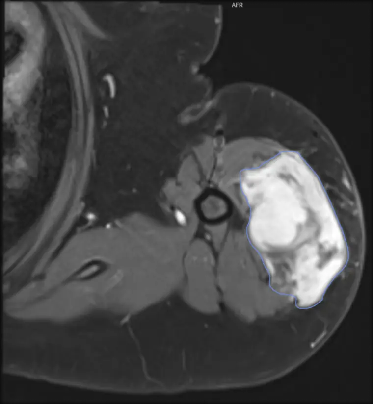
Soft tissue lesion in the arm outlined as a single polygon on a contrast-enhanced slice.
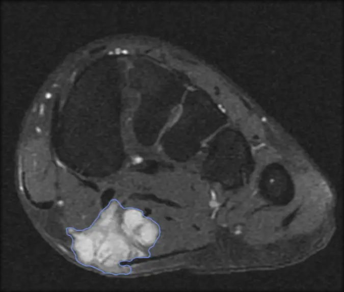
Lesion in the plantar soft tissue outlined on an axial slice of the foot.
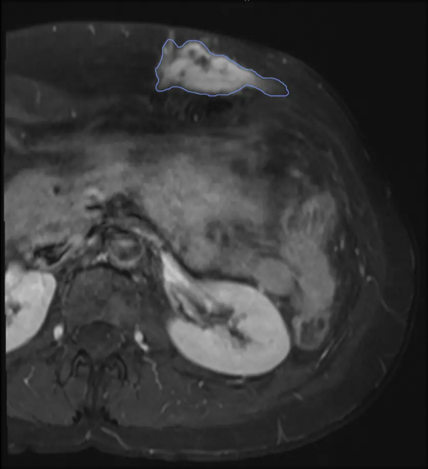
Thin superficial lesion traced along its full length in the abdominal wall.
Chest wall lesion outlined where it sits against the underlying muscle.
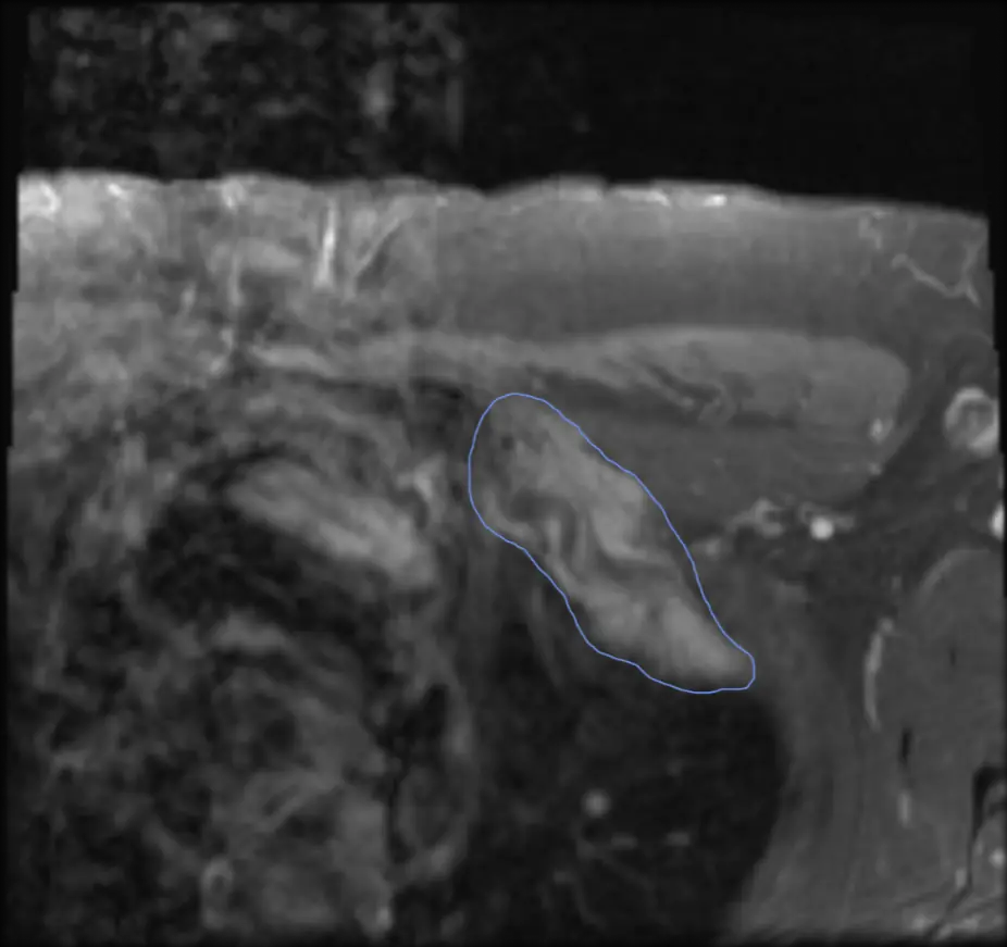
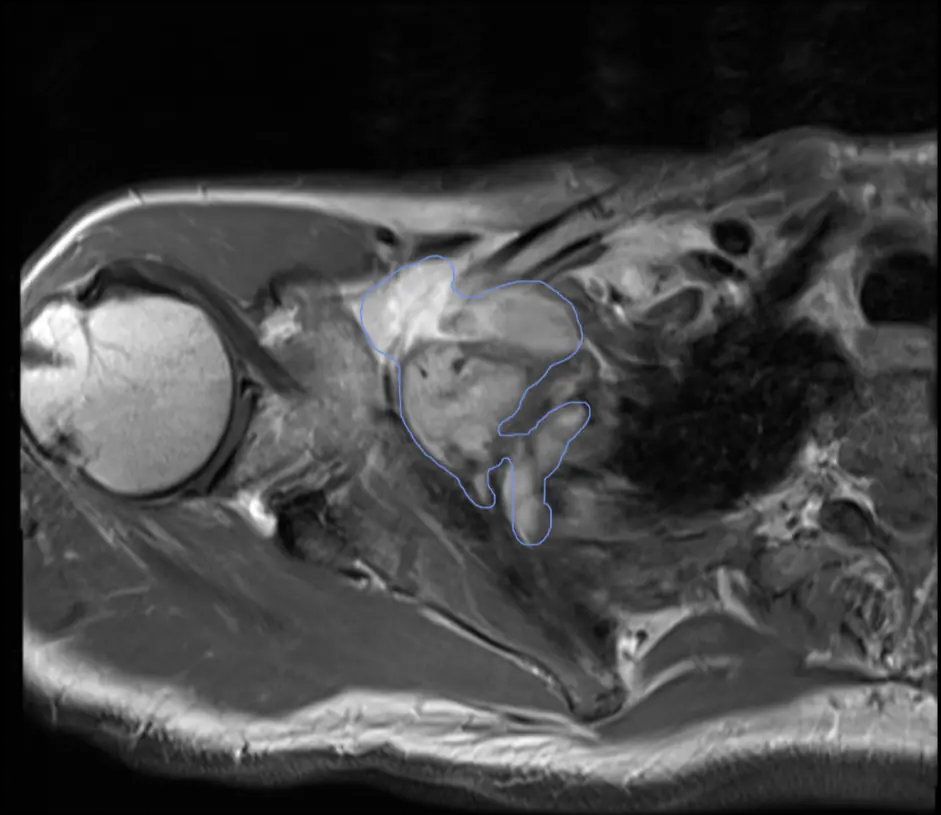
Axillary lesion outlined as one region, including the narrow extension running down from the main body.
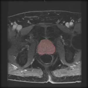
Prostate segmented as a single region on an axial pelvic slice.
Lesions localized and segmented across the head and neck, chest, abdomen and pelvis.
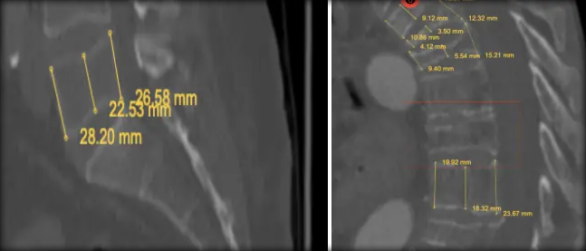
Vertebral levels tagged and inter-level distances measured on sagittal and coronal reformats of one study.
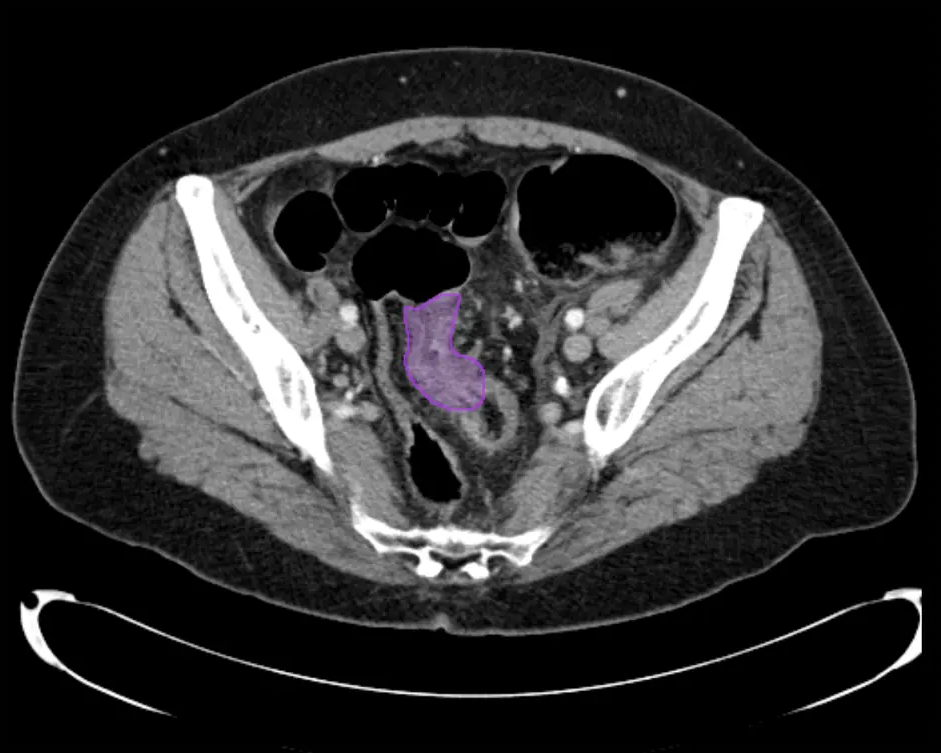
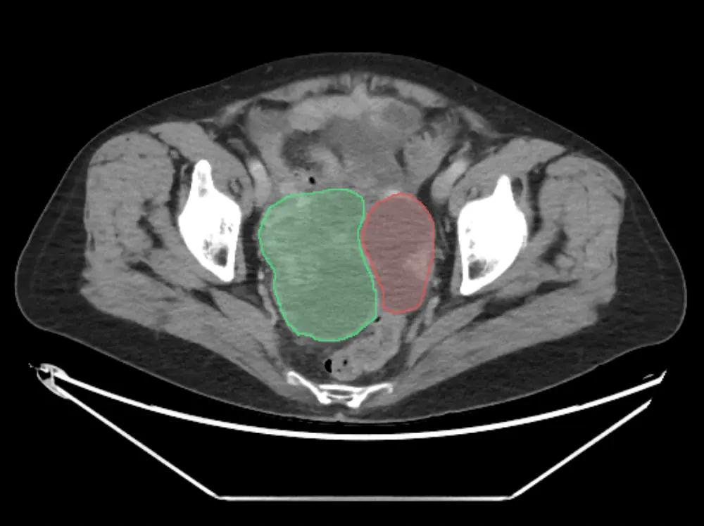
Bowel lesion segmented on an axial pelvic slice.
Ovarian lesion segmented with the adjacent structure labeled as a separate class rather than merged into it.
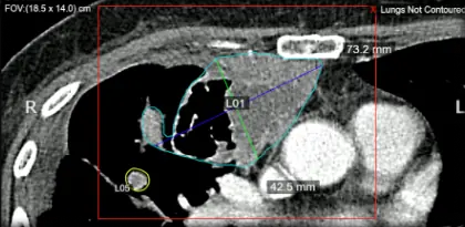
Two lung lesions contoured and labeled separately, each carrying its own axis measurements.
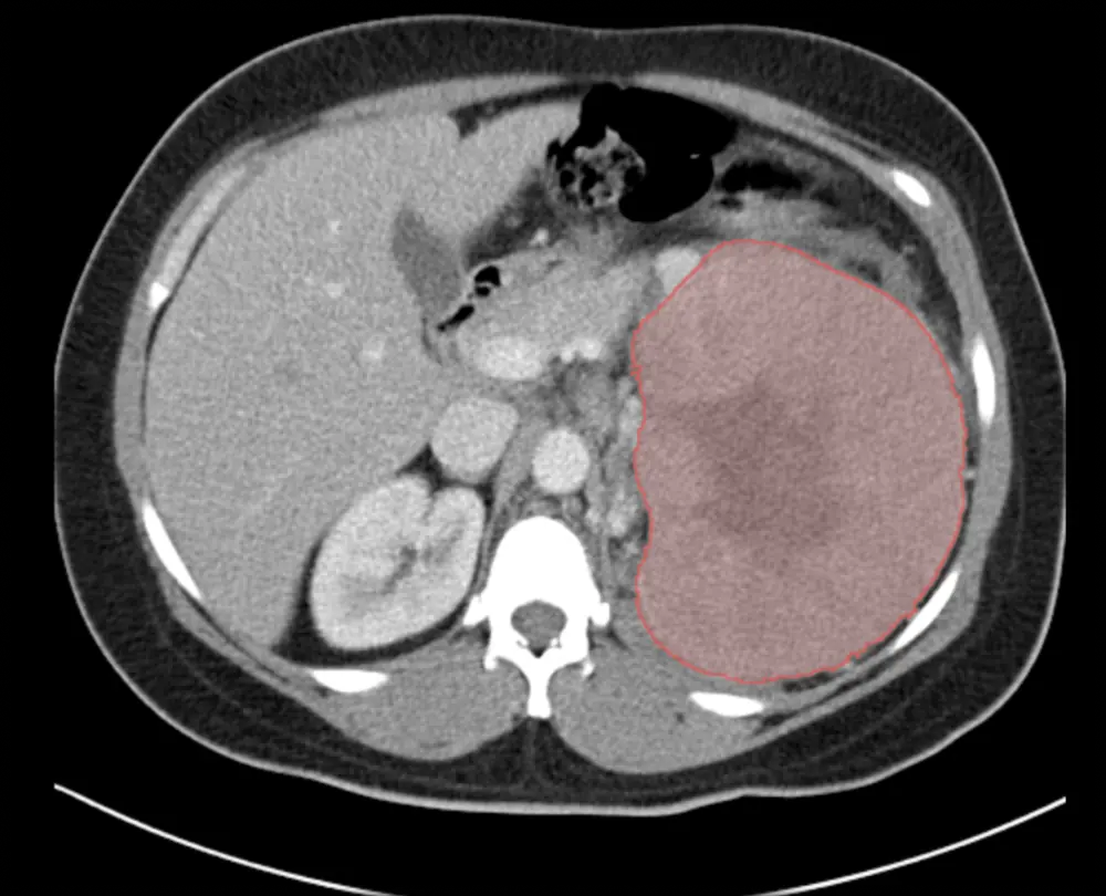
Renal mass segmented as a single region on a contrast-enhanced axial slice.
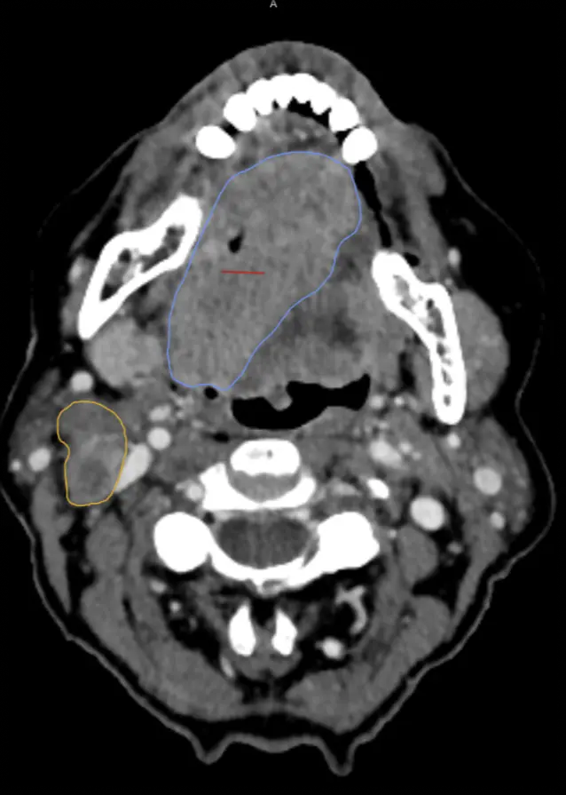
Tongue lesion outlined with a separate cervical structure contoured as its own class.
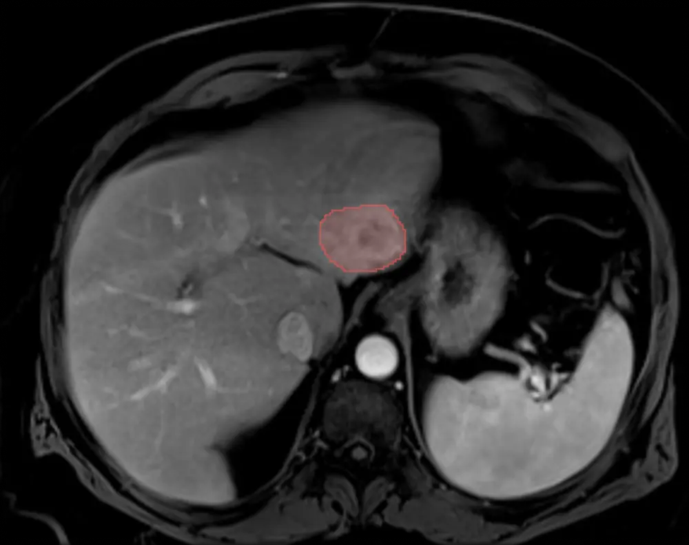
Liver lesion segmented on a contrast-enhanced axial slice.
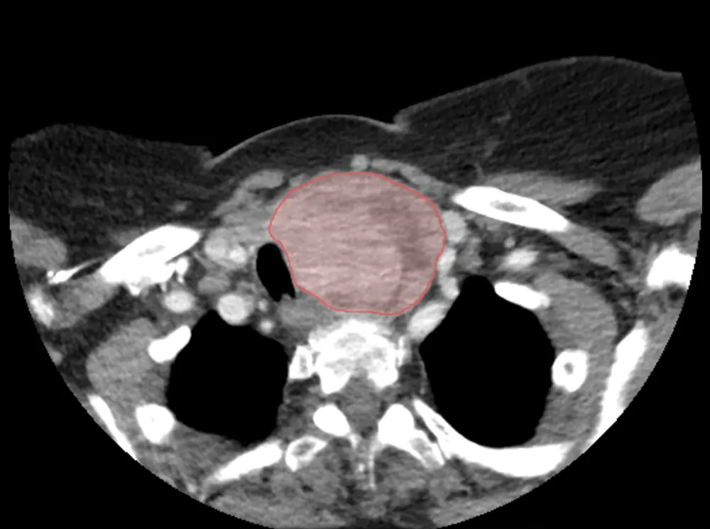
Thyroid lesion segmented on an axial neck slice.
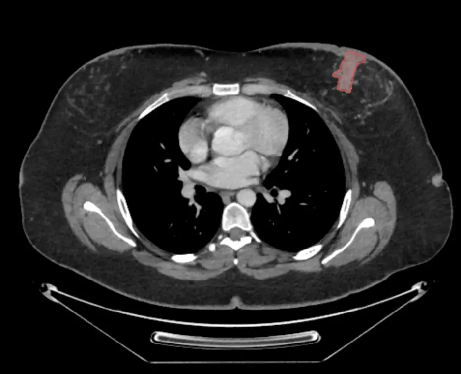
Breast lesion segmented on an axial chest slice.
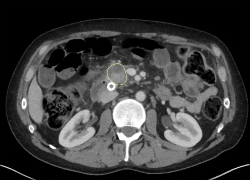
Pancreatic lesion contoured on an axial abdominal slice.
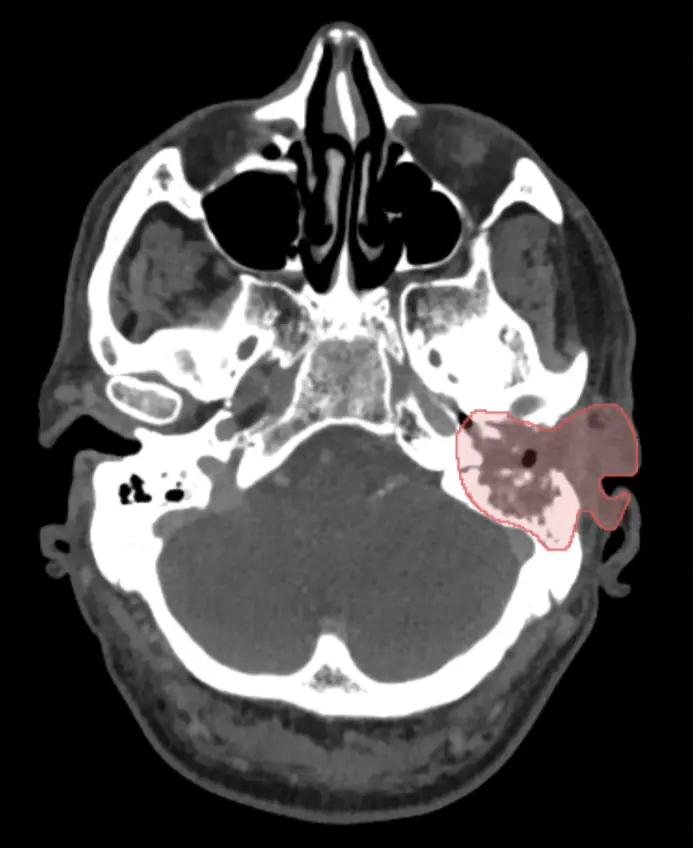
Mastoid lesion segmented on an axial slice through the temporal bone.
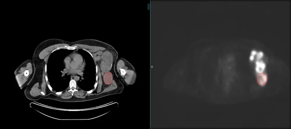
Uptake carried across paired series, and radiographic measurements taken on plain film.
Axillary lesion segmented on the CT slice and carried across to the paired PET image, so the same region can be read on both.
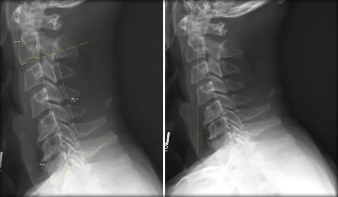
Cobb angle and sagittal vertical axis measured on two lateral spine films.
Masses, calcifications and asymmetries located and labeled, including BI-RADS scoring.
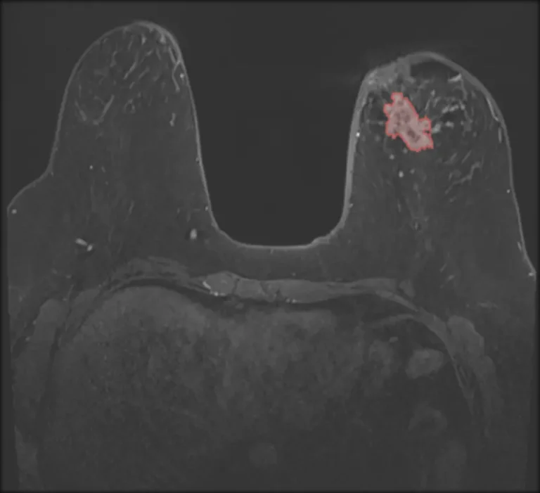
Lesion in the left breast segmented as a single region on an axial slice.
Uptake carried across paired series, and radiographic measurements taken on plain film.
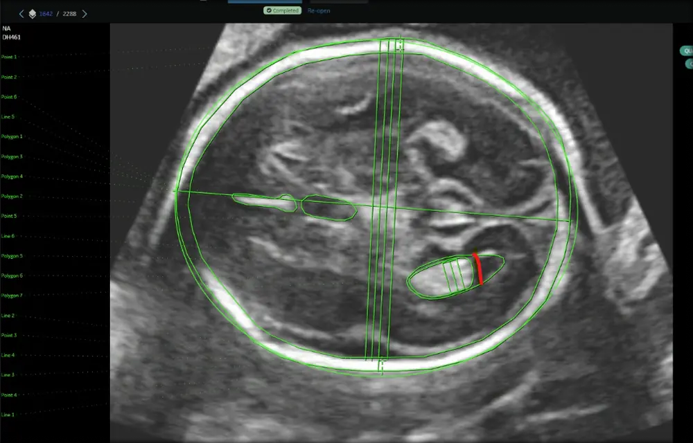
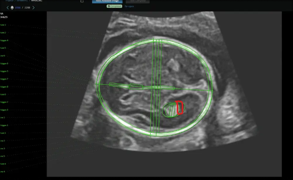
Standard fetal head plane with the circumference ellipse fitted, the axis calipers placed and a midline structure measured separately.
Standard planes measured and cardiac chambers segmented across 2D B-mode, Doppler and cine-loop studies.
Four chamber view segmented into left and right ventricle and left and right atrium, at end diastole and end systole from the same clip.
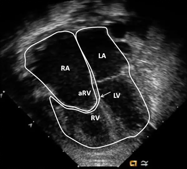
Chamber boundaries traced and labeled on a four chamber view, with the ventricles separated from the atria.
Preoperative and intraoperative studies annotated for real-time guidance, implant placement and minimally invasive procedures.
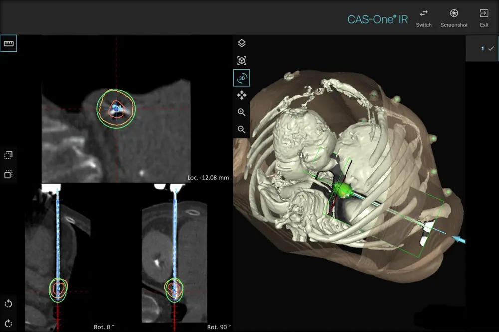
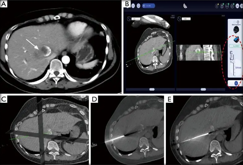
Needle trajectory and target planned across orthogonal reformats and a 3D reconstruction from the same CT study.
Ablation target and instrument path tracked from planning through to the intraprocedural slices.
Annotation of the aortic, mitral, tricuspid and pulmonary valves, including the anatomy of the valves, annulus, leaflets, chambers and vessels, with segmentation, landmarks and measurements for valve planning and interventional procedures.
Modality coverage runs across echocardiography, angiography and cardiac CT and MRI, from coronary vessel labeling on a volume rendered angiogram through to device position on a live fluoroscopic run.
On the electrical side we annotate ECG waveforms, intervals and rhythms, along with EP mapping data covering electrical activation, ablation sites, catheters and anatomical landmarks, for arrhythmia detection and electrophysiology procedures.
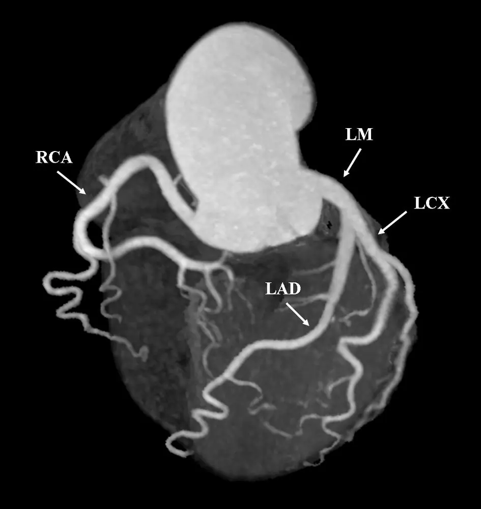
IMAGE TYPES Coronary CT angiography, echocardiography, fluoroscopy, cardiac CT and MRI, ECG and EP mapping
ANNOTATION TYPES Segmentation, landmarks, measurements, waveform and interval labeling
SUPPORTS Valve planning, interventional procedures, arrhythmia detection
Valve and vessel anatomy segmented, landmarked and measured for planning and for procedures in progress.
Left main, left anterior descending, left circumflex and right coronary artery each identified on a volume rendered coronary angiogram.
One valve study read across fluoroscopy, biplane echo views and a 3D reconstruction, with the target annotated on each view.
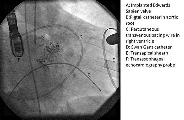
Implanted valve position marked on a fluoroscopic frame, with the delivery wire and sternal wires both visible.
Implanted valve, aortic root catheter, pacing wire, Swan-Ganz catheter, transapical sheath and echo probe each labeled on a single frame.
Waveforms, intervals and rhythms labeled, with EP mapping for activation, ablation sites and catheter position.
The scheme used for ECG labeling: P wave, PR interval and PR segment, QRS complex, ST segment, T wave and QT interval, each marked as its own class.
Anatomical structure identification, surgical instrument detection and tracking, tissue segmentation, surgical phase recognition and procedure assessment.
Anatomy is outlined class by class on both laparoscopic and robotic frames, covering the liver, gallbladder, cystic artery, cystic duct and prostate.
Instruments are located, classified and tracked frame to frame, and procedure stages are recognized and assessed across a full operation.
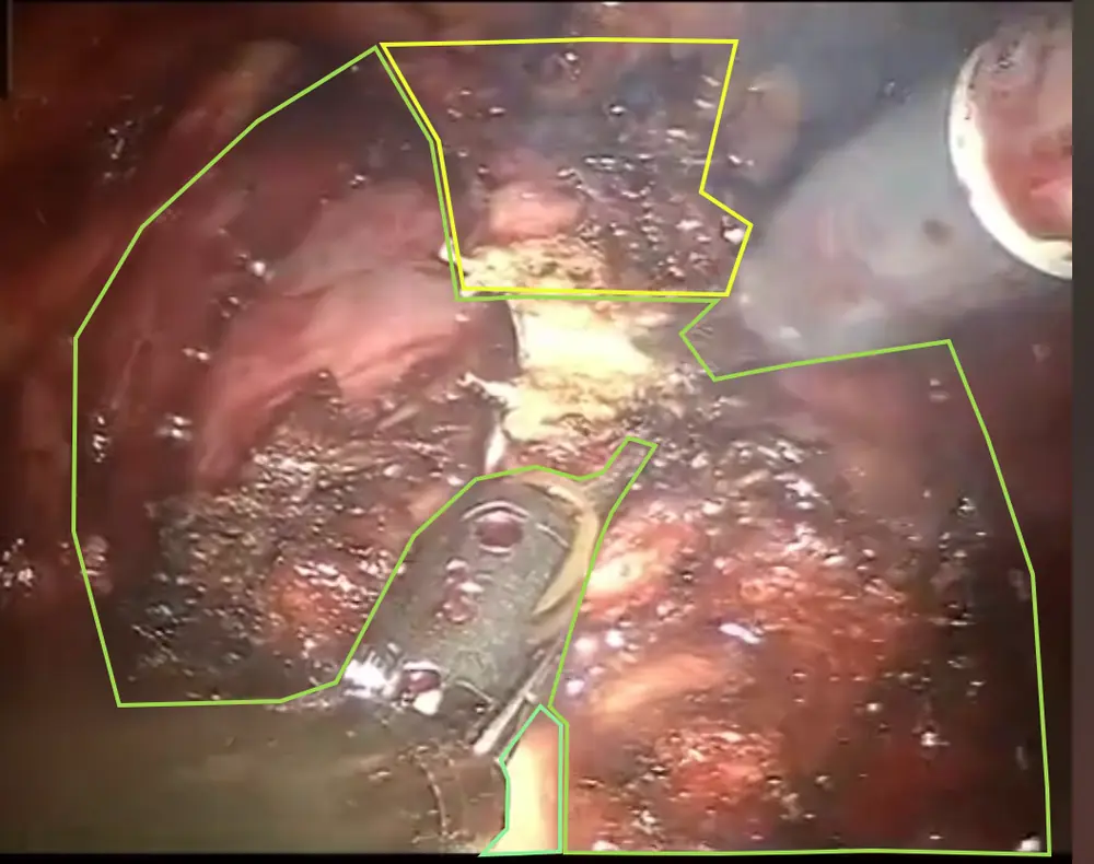
IMAGE TYPES Laparoscopic video, robotic surgical video, still surgical frames
ANNOTATION TYPES Segmentation, polygons, bounding boxes, keypoints, classification, frame-to-frame tracking
SUPPORTS Instrument detection, surgical phase recognition, procedure assessment
Anatomy outlined class by class on laparoscopic and robotic surgical frames.
Prostate and the surrounding tissue planes outlined as separate polygon classes on a robotic laparoscopy frame.
The same anatomy outlined with the polygon vertices visible, showing how the boundary is placed and carried frame to frame.
Liver, gallbladder and the cystic artery and duct outlined as four separate classes in the hepatobiliary triangle.
A second frame from the same procedure, with the instrument in view and the same four classes maintained.
Tools located, classified and tracked across the procedure.
Surgical tool located with a bounding box, and keypoints placed along the shaft and jaws so the instrument can be tracked as it moves.
Artificial Intelligence in Medical Imaging
We assist healthcare and medical AI organizations in building a data and validation foundation to develop reliable, clinically meaningful imaging AI.
Medical Image Annotation
- Image segmentation and classification
- Lesion and anatomical labeling
- Landmark identification and measurements
Clinical Data Preparation
- Medical image curation
- DICOM processing
AI Model Validation
- Clinical and ground-truth validation
- Model performance evaluation
- Edge-case and error analysis
- Bias assessment and inter-rater agreement
- AI output and annotation quality review
AI-assisted Imaging
- Automated disease and lesion detection
- Anatomical segmentation
- Image quality assessment
- Disease severity and risk stratification
- Quantitative imaging and longitudinal analysis
- Clinical decision support and report intelligence
Radiology
MRI, CT, PET-CT, Ultrasound, Mammography, X-ray
Ophthalmology
OCT, Fundus Photography, OCTA, FFA, Anterior Segment OCT
Applications
Oncology, Cardiovascular Imaging, Surgical Imaging, Digital Pathology, Veterinary Imaging
Radiology
MRI, CT, PET-CT, Ultrasound, Mammography, X-ray
Ophthalmology
OCT, Fundus Photography, OCTA, FFA, Anterior Segment OCT
Applications
Oncology, Cardiovascular Imaging, Surgical Imaging, Digital Pathology, Veterinary Imaging
Medical image annotation
- Image segmentation and classification
- Lesion and anatomical labeling
- Landmark identification and measurements
Clinical data preparation
- Medical image curation
- DICOM processing
AI model validation
- Clinical and ground-truth validation
- Model performance evaluation
- Edge-case and error analysis
- Bias assessment and inter-rater agreement
- AI output and annotation quality review
AI-assisted imaging
- Automated disease and lesion detection
- Anatomical segmentation
- Image quality assessment
- Disease severity and risk stratification
- Quantitative imaging and longitudinal analysis
- Clinical decision support and report intelligence
Radiology
CT, MRI, X-ray, ultrasound, PET/CT, mammography
Ophthalmology
OCT, fundus photography, OCTA, FFA, anterior segment OCT
Applications
Oncology, cardiovascular imaging, surgical imaging, digital pathology, veterinary imaging
Image Annotation
- Bounding box: outline objects in images and video for object detection and computer vision.
- Polygon annotation: use multi-vertex contour tracing to preserve the exact shape of irregular objects.
- Key point annotation: mark key points for object pose estimation and motion tracking.
Segmentation and Masking
- Semantic segmentation: classify and label every pixel for high-quality segmentation datasets.
- Image masking: isolate objects, people, and backgrounds with detailed masks.
- Circle and ellipse annotation: label circular and elliptical objects with precise boundaries.
Text and Document Annotation
- Text classification: label intent, topics, and sentiment across text datasets.
- Named entity recognition: tag entities and domain-specific terminology in text.
- Document annotation: identify and label fields and regions across invoices, contracts, forms, and scanned business documents.
Video and Audio Annotation
- Video annotation: label and track objects, actions, and events across video frames.
- Video transcription and labeling: transcribe and label speech, events, actions, and timestamps.
- Audio transcription and labeling: transcribe and label speech, speakers, sounds, and key audio events.
Human expertise and AI
Medical AI requires more than algorithms. Our clinical and technical teams bring expertise in anatomy, pathology, imaging protocols, annotation workflows, quality control and AI validation to every project.
From Sample to Signed-Off Dataset
01
Define
Understand the clinical objective and the AI use case.
02
Curate
Prepare and structure high-quality imaging data.
03
Annotate
Apply clinically relevant labels, measurements and segmentations.
04
Validate
Perform quality and clinical review.
05
Deliver
Provide structured, AI-ready datasets and validation outputs.
Human Expertise and AI
Medical AI requires more than algorithms. Our clinical and technical teams bring expertise in anatomy, pathology, imaging protocols, annotation workflows, quality control and AI validation to every project.
-
Define
Understand the clinical objective and the AI use case. -
Curate
Prepare and structure high-quality imaging data. -
Annotate
Apply clinically relevant labels, measurements and segmentations. -
Validate
Perform quality and clinical review. -
Deliver
Provide structured, AI-ready datasets and validation outputs.
Ready to Get Your Data Labeled?
Share a sample and your requirements. We will prepare a sample annotation, so you can review the quality before moving forward.
Talk to Our Experts
Tell us what you’re trying to achieve, and we’ll get back to you.